


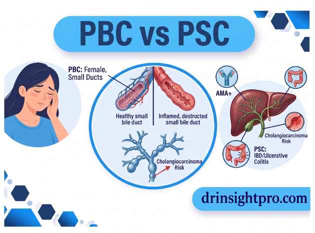
Patients often hear these two similar-sounding names [PBC vs PSC] together and wonder whether they are actually the same disease. They are not. Both primary biliary cholangitis (PBC) vs primary sclerosing cholangitis (PSC) damage the bile ducts, which carry bile from the liver to the intestine. However, they affect different parts of the biliary system, occur in different patient groups and require different management strategies.
This guide explains both conditions in plain language. It also shows how doctors distinguish PBC vs PSC when routine blood tests first raise suspicion.
PBC vs PSC: The Key Difference in One Minute
PBC mainly targets the small bile ducts inside the liver, and most patients with PBC have antimitochondrial antibodies (AMA). In contrast, PSC causes inflammation and scarring of the bile ducts and can involve ducts inside the liver, outside the liver or both.
Doctors usually treat PBC first with ursodeoxycholic acid (UDCA), which provides established disease-modifying benefits. However, no medication currently prevents or reverses PSC progression.Therefore, PSC management focuses on monitoring, treating complications and considering liver transplantation when appropriate.
PBC vs PSC: Quick Comparison
| Feature | Primary Biliary Cholangitis (PBC) | Primary Sclerosing Cholangitis (PSC) |
|---|---|---|
| Typical sex affected | Women | Men |
| Typical age at diagnosis | Middle age, often 40–60 | Often younger to middle-aged adults |
| Main process | Immune-mediated destruction of small bile ducts | Fibroinflammatory scarring and narrowing of bile ducts |
| Bile ducts involved | Small intrahepatic ducts | Intrahepatic ducts, extrahepatic ducts or both |
| IBD association | Not characteristic | Strong association, especially with ulcerative colitis |
| Antimitochondrial antibody | Usually positive | Usually negative |
| Role of MRCP | Usually unnecessary when diagnostic criteria are already met | Preferred imaging test for suspected large-duct PSC |
| Small-duct disease | Characteristic of PBC | Small-duct PSC occurs when large ducts appear normal |
| Biliary strictures | Not a typical feature | Important complication |
| Cholangiocarcinoma risk | Not a defining risk | Important concern |
| Colorectal cancer risk | Not characteristically increased | Increased, particularly with coexisting IBD |
| First-line treatment | UDCA | No approved disease-modifying drug |
| Advanced disease | Liver transplantation when indicated | Liver transplantation when indicated |
Both conditions can recur after liver transplantation in some patients. Nevertheless, transplantation remains the definitive treatment for appropriately selected patients with advanced disease.
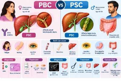
What Do PBC and PSC Have in Common?
Despite their differences, both conditions can cause:
- Cholestatic liver test abnormalities, particularly an elevated alkaline phosphatase (ALP)
- Fatigue
- Pruritus or itching
- Jaundice in more advanced disease
- Fat-soluble vitamin deficiency when cholestasis becomes significant
- Increased osteoporosis risk
- Cirrhosis and liver failure in some patients
- A need for ongoing specialist follow-up
What Is Primary Biliary Cholangitis (PBC)?
In PBC, the immune system attacks and damages the small bile ducts inside the liver. As a result, bile flow becomes impaired and cholestasis develops.
Doctors previously called the condition primary biliary cirrhosis. However, doctors changed the name because many patients receive the diagnosis before cirrhosis develops.
PBC can also cause an abnormal lipid profile. Therefore, doctors should assess cardiovascular risk according to the patient’s overall risk factors rather than interpreting the lipid profile in isolation.
If PBC progresses to cirrhosis, the risk of hepatocellular carcinoma (HCC) increases. Therefore, patients with cirrhosis require appropriate HCC surveillance.
Who Gets PBC?
PBC predominantly affects women and commonly appears between 40 and 60 years of age. A close family history can increase individual risk.
Furthermore, PBC can occur alongside autoimmune conditions such as Sjögren syndrome, autoimmune thyroid disease and celiac disease. Doctors therefore assess patients for these conditions according to their symptoms and clinical history.
Symptoms of PBC
Many patients have no symptoms during the early stages. When symptoms develop, they may include:
- Persistent fatigue
- Itching, particularly at night
- Dry eyes and dry mouth
- Xanthelasma or xanthomas
- Jaundice in more advanced disease
Importantly, the severity of fatigue or itching does not always reflect the stage of liver disease.
How Is PBC Diagnosed?
Doctors generally diagnose PBC when at least two of the following three criteria are present:
- A cholestatic biochemical profile, typically with an elevated ALP
- PBC-specific autoantibodies, especially AMA
- Compatible liver biopsy findings
Therefore, a patient with cholestatic liver tests and a positive AMA usually does not need a biopsy to establish the diagnosis.
However, when PBC-specific antibodies are negative or the diagnosis remains uncertain, liver biopsy may help establish the diagnosis and assess other possible causes of cholestasis.
How Is PBC Treated?
PBC treatment generally follows a structured approach:
- Start UDCA as first-line disease-modifying treatment.
- Assess the biochemical response after approximately 6–12 months.
- Consider an approved second-line therapy if the response is inadequate or the patient cannot tolerate UDCA.
- Treat pruritus separately when necessary.
- Assess fibrosis or cirrhosis and monitor for complications.
- Consider liver transplantation when advanced disease warrants it.
Newer second-line options include the PPAR agonists elafibranor (Iqirvo) and seladelpar (Livdelzi). Both received accelerated FDA approval in 2024 for adults with PBC who have an inadequate response to UDCA or cannot tolerate it.
These approvals were based primarily on biochemical endpoints. Therefore, longer-term clinical outcome data will continue to provide important information about their effects on disease progression.
Obeticholic acid was previously used as a second-line option. However, its regulatory status and use have changed because of safety concerns. Therefore, it should not be presented as equivalent to the currently available second-line options.
What Is Primary Sclerosing Cholangitis (PSC)?
PSC is a chronic fibroinflammatory disease that causes inflammation, scarring and narrowing of the bile ducts. It can affect the large ducts, small ducts or both.
The scarring creates alternating areas of narrowing and dilation. As a result, imaging may show the characteristic beaded appearance of the biliary tree.
Who Gets PSC?
PSC commonly affects men and often develops in younger or middle-aged adults. One of its most important features is its strong association with inflammatory bowel disease (IBD), particularly ulcerative colitis.
Therefore, clinicians should consider PSC when a patient with IBD develops unexplained cholestatic liver test abnormalities.
Symptoms of PSC
PSC can remain silent for years and may first appear through abnormal liver tests. When symptoms develop, they can include:
- Fatigue
- Itching
- Jaundice
- Poor appetite
- Abdominal or digestive discomfort
- Weight loss in advanced disease
Because impaired bile flow affects digestion and nutrient absorption, significant cholestasis can also lead to fat-soluble vitamin deficiencies and bone disease.
How Is PSC Diagnosed?
MRCP is the preferred non-invasive imaging test for suspected large-duct PSC. It provides detailed images of the biliary system and can show multifocal strictures and areas of dilation.
In classic PSC, these changes can create a characteristic beaded appearance.
Unlike PBC, PSC has no single blood antibody that confirms the diagnosis. Perinuclear ANCA can be positive in many patients, but it lacks sufficient specificity to establish PSC.
What Is Small-Duct PSC?
Some patients have clinical and biochemical features suggestive of PSC even though their large bile ducts appear normal on MRCP.
In this situation, liver biopsy may help establish small-duct PSC after other causes have been considered. Small-duct PSC generally has a more favorable course than large-duct PSC. However, some patients later develop large-duct involvement.
How Is PSC Treated?
Currently, no medication is approved specifically to prevent or reverse PSC progression. Therefore, treatment focuses on monitoring the disease and managing its complications.
Management may include:
- ERCP: Used mainly for clinically significant biliary strictures, particularly when cholangitis, worsening cholestasis or suspected malignancy is present. Balloon dilation is generally preferred while stenting is reserved for selected situations.
- Antibiotics: Used to treat bacterial cholangitis.
- IBD surveillance: Patients with PSC and IBD require appropriate colorectal cancer surveillance.
- Liver transplantation: Remains the definitive treatment for advanced PSC.
UDCA can improve liver biochemical tests in some patients with PSC. However, improved biochemical results have not been shown to translate into improved clinical outcomes. Therefore, routine use solely to treat PSC is not generally recommended.
How Do Doctors Tell PBC vs PSC Apart?
When cholestatic liver tests raise suspicion for a bile duct disorder, doctors usually follow a step-by-step approach.
- Confirm that the elevated ALP originates from the liver or biliary system rather than another source such as bone.
- Use abdominal ultrasound to look for mechanical biliary obstruction.
- Test for AMA when PBC is suspected.
- A positive AMA together with compatible cholestatic liver tests strongly supports PBC.
- If AMA is negative or PSC is suspected, particularly in a patient with IBD, perform MRCP.
- If MRCP is normal but suspicion remains high, consider small-duct PSC or another cholestatic disorder. Liver biopsy may then be appropriate.
| Finding | What it may suggest |
|---|---|
| High ALP with positive AMA | PBC |
| High ALP with IBD | Consider PSC |
| MRCP showing multifocal strictures and dilation | PSC |
| Normal MRCP with persistent suspicion | Consider small-duct PSC or another cholestatic condition |
| Biliary obstruction on ultrasound | Consider mechanical obstruction such as a gallstone |
PBC vs PSC: Pathology Differences
| Feature | PBC | PSC |
|---|---|---|
| Main target | Small intrahepatic bile ducts | Intrahepatic ducts, extrahepatic ducts or both |
| Characteristic process | Immune-mediated destruction of small ducts | Fibroinflammatory stricturing |
| Classic histologic clue | Florid duct lesion | Periductal fibrosis, sometimes described as an onion-skin pattern |
PBC vs PSC: Complications and Cancer Risk
Both diseases require long-term monitoring. However, the major cancer risks differ.
In PBC, the risk of HCC becomes particularly important when cirrhosis develops. Therefore, patients with PBC-related cirrhosis should follow appropriate HCC surveillance recommendations.
In PSC, cholangiocarcinoma is an important concern. PSC also increases the risk of gallbladder cancer and colorectal cancer in patients who also have IBD.
Surveillance strategies vary between guidelines and individual risk factors. Depending on the clinical situation, monitoring may include MRI/MRCP, other imaging or CA 19-9 testing. However, CA 19-9 alone cannot diagnose cholangiocarcinoma because it can increase for reasons unrelated to cancer.
Bone health also deserves attention in both conditions. Chronic cholestasis can contribute to vitamin deficiencies and osteoporosis. Therefore, doctors should assess bone health according to disease duration and individual risk factors.
Two Composite Patient Scenarios
The following fictional scenarios illustrate typical diagnostic pathways. They do not describe individual patients.
Scenario 1: A 52-year-old woman with fatigue and itching
She develops persistent fatigue and nighttime itching. Routine blood tests show a cholestatic pattern with an elevated ALP. AMA is strongly positive and abdominal ultrasound shows no biliary obstruction.
Because she meets the clinical and laboratory criteria for PBC, a biopsy is not required to establish the diagnosis. She starts UDCA and undergoes follow-up testing to assess her biochemical response.
Scenario 2: A 29-year-old man with ulcerative colitis
He has a five-year history of ulcerative colitis. Although his bowel symptoms remain controlled, routine liver testing reveals an elevated ALP.
Because PSC can develop silently in patients with IBD, his doctor orders MRCP. The scan shows multifocal bile duct strictures and dilation consistent with large-duct PSC.
He then begins an individualized monitoring plan that addresses both PSC and his underlying IBD.
These cases highlight an important difference: PBC diagnosis often relies heavily on AMA testing whereas PSC diagnosis relies primarily on biliary imaging.
          Elevated ALP ± GGT
                 │
                 ▼
   Confirm hepatic/biliary origin
                 │
                 ▼
   Abdominal Ultrasound
   Assess for biliary obstruction
                 │
      ┌──────────┴──────────┐
      ▼                     ▼
 No obstruction       Obstruction present
      │                     │
      ▼                     ▼
    Check AMA          Investigate and
      │                treat obstruction
 ┌────┴────┐
 ▼         ▼
AMA Positive AMA Negative
│ │
▼ ▼
Compatible Consider PSC,
cholestatic other cholestatic
pattern liver diseases
│ │
▼ ▼
PBC MRCP if PSC
is suspected
│
┌──────┴──────┐
▼ ▼
Multifocal Normal
strictures large ducts
/ beading │
│ ▼
▼ Persistent suspicion
PSC of small-duct PSC
│
▼
Consider liver biopsy
when appropriate
PBC vs PSC: Recent Research and New Treatments
PBC treatment has expanded considerably in recent years. Elafibranor and seladelpar received accelerated FDA approval in 2024 for adults with PBC who have an inadequate response to UDCA or cannot tolerate it.
In the pivotal phase 3 trial of seladelpar, 62% of participants achieved the specified composite biochemical response at 12 months compared with 20% receiving placebo. Seladelpar also reduced itching in patients with PBC.
However, these approvals rely primarily on biochemical outcomes. Therefore, longer-term evidence will help clarify their effects on major clinical outcomes.
PSC research continues to explore therapies that could modify disease progression. For example, clinical trials have investigated agents such as cilofexor and norursodeoxycholic acid. However, these investigational therapies are not currently approved as routine disease-modifying treatments for PSC.
PBC vs PSC: What Are We Still Learning?
Research continues to explore several important questions.
- Fatigue: PBC-related fatigue can significantly affect quality of life and does not always correspond to liver disease severity.
- Bone health: Both PBC and PSC can increase osteoporosis risk. Therefore, early assessment of individual bone health is important.
- Pregnancy: Many patients with PBC or PSC can become pregnant. However, disease activity, liver function, itching and medication safety require individualized planning with hepatology and obstetric teams.
When Should You See a Doctor?
You do not need to wait for jaundice before getting checked.
See a doctor if you develop:
- Persistent unexplained fatigue
- Persistent itching without a rash
- Yellowing of the skin or eyes
- Unexplained abnormal liver tests, particularly an elevated ALP
If you have IBD, regular liver testing can help identify PSC even when you have no symptoms.
Frequently Asked Questions
No, They are distinct diseases with different underlying mechanisms. Rare cases may show overlapping features, but one disease does not simply transform into the other.
Currently, no approved medication has been shown to prevent or reverse the natural progression of PSC. Liver transplantation remains the definitive treatment for advanced disease.
PSC. Its association with IBD, particularly ulcerative colitis, is one of its most important clinical features.
Medical Disclaimer
This article is intended for general educational purposes and does not replace professional medical advice, diagnosis or treatment. Each patient’s situation is different. Therefore, decisions about testing and treatment should be made with a qualified healthcare professional who understands the patient’s medical history.
About the Author
This article was written by Hafsa Hadi, a health content writer focused on making liver and digestive disease information clear and accessible for general readers. This article should undergo review by a licensed gastroenterologist before publication.
References
AASLD Practice Guidance on Primary Biliary Cholangitis (2018 / 2021 Update):
- Full Guidance PDF: AASLD PBC Practice Guidance
- PubMed Citation: PubMed: AASLD 2021 Guidance Update
- Key topics covered: Diagnostic criteria (ALP $\ge 1.5\times$ ULN, positive AMA, histology), weight-based UDCA dosing (13–15 mg/kg/day), and updated safety advisories on obeticholic acid in decompensated cirrhosis.
AASLD Practice Guidance on Primary Sclerosing Cholangitis and Cholangiocarcinoma (2023):
- Full Guidance PDF: AASLD PSC & CCA Practice Guidance
- PubMed Citation: PubMed: Bowlus et al., Hepatology 2023
- Key topics covered: MRCP as first-line imaging, dominant stricture management, avoidance of diagnostic ERCP, and colorectal/cholangiocarcinoma surveillance protocols.
NIH / NIDDK Patient and Clinical Education:
- NIDDK: Primary Biliary Cholangitis Overview
- NIDDK: Primary Sclerosing Cholangitis Overview
- Useful for accessible, plain-language patient education and background pathophysiology.
FDA Approvals for Second-Line PBC Therapies:
- Elafibranor (Iqirvo): FDA Drug Information & Labeling (accelerated approval for UDCA-inadequate responders)
- Seladelpar (Livdelzi): FDA Drug Approval History (selective PPAR $\delta$ agonist approved for biochemical response and pruritus relief)