


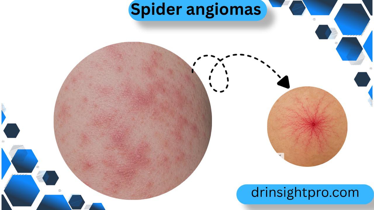
Spider angiomas is a small vascular mark with a central red spot and thin vessels spreading outward like a web. A single mark or a small number can appear naturally in healthy children, young adults and pregnant women. Moreover, multiple marks appearing together, especially alongside signs like jaundice or abdominal swelling, can signal chronic liver disease and warrant medical evaluation.
What Is a Spider Angiomas?
A spider angioma is an abnormal collection of small dilated blood vessels located directly beneath the surface of the skin. In addition, medical professionals also call this lesion a spider nevus, spider telangiectasia or vascular spider. It derives its name from its distinct visual shape. However, the lesion has three defining anatomical features:
- Central Arteriole: A tiny elevated red dot at the center that acts as the primary feeding vessel supplying blood to the rest of the mark.
- Radiating Capillaries: Thin branching blood vessels that travel outward in all directions from the central core like the legs of a spider.
- Rapid Blanching Response: Pressing gently on the center of the lesion with a fingertip or a clear glass slide empties the vessels, causing the entire mark to turn pale or match the surrounding skin tone. Releasing the pressure allows blood to refill the vessels rapidly from the center outward within one to two seconds. This rapid refill pattern confirms that the lesion is fed by an arteriole and helps doctors separate it from other red skin spots.
Where Do Spider Angiomas Appear on the body?
Furthermore, spider angiomas almost exclusively appear on areas of the body that correspond to the superior vena cava distribution. This pattern includes the upper half of the body:
- Face and forehead
- Neck and throat
- Upper chest and collarbone area
- Upper back and shoulders
- Forearms, wrists and backs of the hands
In addition, spider angiomas rarely form on the lower abdomen, legs or feet.

Spider Angiomas vs Other Red Skin Marks: key Differences
Many vascular and dermatological conditions produce red marks on the skin. However, a careful physical evaluation helps distinguish them accurately:
| Skin Condition | Primary Biological Cause | Typical Visual Appearance | Blanching Test Result | Common Body Locations |
| Spider Angioma | Dilated central feeding arteriole with branching vessels | Red central dot with radiating thin capillary legs | Blanches fully and refills rapidly from the center | Face, neck, upper chest, shoulders and arms |
| Cherry Angioma | Benign overgrowth of mature skin capillaries | Smooth round bright red or purple dome-shaped bump | Does not blanch or only fades slightly | Chest, back, abdomen and limbs |
| Spider Veins | Dilated venules caused by high local venous pressure | Web-like or jagged purple and blue linear lines | Minimal blanching with slow refill | Thighs, calves and ankles |
| Petechiae | Microscopic bleeding from broken capillaries under skin | Tiny flat pinpoint red, brown or purple dots | Does not blanch at all under pressure | Lower legs, ankles, arms and mucous membranes |
| Telangiectasia | Permanently widened superficial blood vessels | Fine red or pink lines without a central feeding dot | Blanches easily but lacks radial refill pattern | Cheeks, nose and around the nostrils |
What Causes Spider Angiomas in liver Disease and Health?
Spider angiomas develop when vascular tone, hormone clearance and vessel growth signals become disrupted.
Chronic Liver Injury or Cirrhosis
       │
       ├──► Reduced hormone clearance ──► Elevated free estrogen levels ──┐
       │                                                                  ├──► Superficial Arteriolar Dilation
       └──► Endothelial growth signals ──► Rise in VEGF and Substance P ──┘    (Spider Angioma Formation)
1. Elevated Estrogen Levels
Moreover, a healthy liver continuously filters, metabolizes and clears steroid hormones from the bloodstream. When liver cells experience progressive damage, hormone clearance slows down, causing free estrogen levels to rise. Therefore, high levels of circulating estrogen relax the smooth muscles lining arterial walls, which prompts small blood vessels near the skin surface to widen permanently.
2. Angiogenic Growth Factors and Neuropeptides
Similarly, chronic liver injury alters endothelial cell signaling. Therefore, the body produces increased levels of vascular endothelial growth factor (VEGF), basic fibroblast growth factor (bFGF) and substance P. Moreover, these biochemical messengers stimulate the proliferation of new microvessels and cause existing capillaries to expand into spider like shapes.
Common vs. Disease-Related Causes
1. Physiologic and Hormonal Causes of Spider Angiomas
- Pregnancy: Surging estrogen levels during the second and third trimesters frequently lead to multiple spider angiomas. However, these marks are harmless and typically disappear on their own within several weeks or months after childbirth as hormone levels return to baseline.
- Healthy Young People: Up to 15% of healthy children, teenagers and young adults develop one or two isolated marks without any internal illness. In addition, these lesions can remain stable or fade with age.
- Exogenous Hormone Use: Birth control pills and hormone replacement therapy can raise estrogen levels enough to stimulate vessel dilation in susceptible individuals.
2. Chronic Liver Disease and Systemic Causes of Spider Angiomas
- Liver Cirrhosis: Approximately one third of patients diagnosed with liver cirrhosis exhibit multiple spider angiomas. Common underlying causes include alcohol associated liver disease, metabolic dysfunction associated steatohepatitis (MASH) and chronic viral hepatitis.
- Chronic Hepatitis B and C: In addition, longstanding viral inflammation of the liver impairs hepatic synthetic and metabolic function over time.
- Thyrotoxicosis: However, an overactive thyroid gland creates a hypermetabolic state with increased peripheral blood circulation that can induce superficial vessel widening.
- Rheumatoid Arthritis: Autoimmune inflammation and associated vascular changes can occasionally cause spider angiomas.
- Severe Malnutrition: Prolonged protein and nutrient deficiencies impair metabolic liver function and hormone clearance.
The Liver Connection and Diagnostic Significance
Medical practitioners pay close attention to the number, pattern and persistence of spider angiomas during a physical exam:
- High Specificity for Chronic Liver Disease: While a single isolated lesion is common in the general public, the presence of multiple spider angiomas carries high clinical specificity for chronic liver disease. Multiple marks indicate that systemic hormone metabolism and vascular regulation are compromised.
- Correlation with Portal Hypertension and Esophageal Varices: Spider angiomas do not develop directly from mechanical backward pressure inside the portal vein system. Instead, portal hypertension, esophageal varices and spider angiomas are all downstream consequences of worsening liver cirrhosis. When a patient presents with prominent spider angiomas, doctors routinely check for portal hypertension and internal varices to prevent dangerous internal bleeding.
Risk Factors for Pathologic Spider Angiomas
You have a higher probability of developing spider angiomas related to underlying liver conditions if you have:
- A history of long term heavy alcohol consumption
- Chronic infection with hepatitis B or hepatitis C virus
- Obesity, type 2 diabetes or metabolic syndrome linked to fatty liver disease
- Autoimmune conditions affecting the liver or joints
- A family history of genetic liver disorders such as hemochromatosis or Wilsons disease
Warning Signs and When to Seek Immediate Medical Care
Mostly, a spider angioma itself does not cause physical pain, itching or skin breakdown. However, the primary concern is the underlying medical condition that triggered it.
Schedule an appointment with a doctor if you notice:
- New or multiplying spider angiomas on your upper body
- Yellowing of your skin or the whites of your eyes (jaundice)
- Unexplained persistent fatigue, daytime drowsiness or muscle weakness
- Swelling in your abdomen (ascites) or fluid buildup in your legs and ankles (edema)
- Unusually red or warm palms (palmar erythema)
- Easy bruising, frequent nosebleeds or slow blood clotting from small cuts
- Unintended weight loss and loss of appetite
Emergency Warning Signs
Seek urgent emergency medical care immediately if you experience:
- Vomiting bright red blood or dark coffee ground material
- Passing black, sticky or foul smelling stools (melena)
- Sudden severe abdominal pain
- Acute mental confusion, slurred speech or severe disorientation (hepatic encephalopathy)
Diagnosis of Spider Angiomas
In addition, a medical workup typically follows a structured step-by-step process:
1. Clinical Examination
Moreover, the doctor inspects the lesion, assesses its location, counts the total number of marks and performs the manual blanch test using light pressure to confirm arteriolar refill.
2. Comprehensive Blood Tests
- Liver Injury Enzymes: Often, Alanine aminotransferase (ALT) and aspartate aminotransferase (AST) evaluate acute or ongoing hepatocellular damage.
- Biliary Enzymes: Also, Alkaline phosphatase (ALP) and gamma glutamyl transferase (GGT) assess bile duct health.
- Hepatic Synthetic Function: Furthermore, Serum albumin, total bilirubin and prothrombin time with international normalized ratio (PT INR) measure the livers capacity to synthesize vital proteins and clear waste.
- Complete Blood Count (CBC): Platelet counts are checked carefully because an enlarged spleen from portal hypertension often traps and lowers circulating platelets.
- Viral Serologies: Additionally, blood panels screen for hepatitis B surface antigen and hepatitis C antibodies.
3. Noninvasive Imaging and Fibrosis Tests
- Abdominal Ultrasound: Likewise, it visualizes liver size, structural contour, surface nodularity, spleen enlargement and blood flow direction in the portal vein.
- Transient Elastography (FibroScan): Likewise, it uses sound waves to measure liver tissue stiffness, providing an accurate score for liver fibrosis and cirrhosis without a biopsy.
4. Endoscopic Screening
- Upper Endoscopy (Esophagogastroduodenoscopy or EGD): If tests confirm cirrhosis or significant portal hypertension, a gastroenterologist uses a thin camera to look directly at the esophagus and stomach for enlarged varices that might require medical banding or preventive medication.
Treatment and Medical Management of Spider Angioma
Treatment depends on whether the marks are linked to an underlying illness or are purely cosmetic:
1. Medical Management of Underlying of Spider Angiomas
When spider angiomas stem from liver disease, treatment focuses entirely on the root cause rather than the skin marks:
- While, total abstinence from alcohol allows liver inflammation to subside.
- Equally, antiviral therapies for chronic hepatitis B or C suppress viral activity and halt progressive liver injury.
- Rather, weight management, healthy diet and blood sugar control reduce liver fat and inflammation in metabolic liver disease.
- Despite this, management of portal hypertension using beta-blocker medications reduces internal pressure.
- Successful liver transplantation in patients with end stage liver failure restores normal hormone metabolism, causing spider angiomas to shrink or disappear completely.
2. Cosmetic Removal Procedures of Spider Angiomas
Conversely, for individuals without liver disease who want marks removed for personal aesthetic reasons, a dermatologist can perform targeted office procedures:
- Pulsed Dye Laser (PDL) Therapy: Delivers concentrated light beams that selectively heat and close the central feeding arteriole without burning surrounding skin tissue.
- Electrocautery and Electrodesiccation: Uses a microscopic needle delivering a tiny electrical current to seal the central blood vessel.
On the other hand, cosmetic treatments only eliminate the visible superficial mark and do not treat or improve any internal liver disorder.
Prevention from Spider Angioma
While you cannot prevent spider angiomas caused by genetics or normal pregnancy, you can protect your liver and lower your risk of chronic liver disease:
- Limit or eliminate alcohol consumption according to clinical recommendations.
- Receive the hepatitis B vaccination series.
- Get screened for hepatitis C if you have potential exposure risk factors.
- Maintain a balanced diet, exercise regularly and manage cholesterol and blood glucose levels to prevent fatty liver disease.
- Attend routine health checkups with your doctor if you take medications that affect the liver.
Frequently Asked Questions
Yes, rising estrogen during pregnancy commonly triggers spider angiomas, especially in the second and third trimesters. Most resolve within weeks to months after delivery as hormone levels return to normal.
Spider angiomas are small clusters near the skin surface with a central feeding vessel, usually found on the upper body. Spider veins are larger and more widespread, commonly found on the legs, and often linked to circulation problems rather than the liver.
No, a spider angioma itself is harmless and painless. The concern arises when several appear together, since that pattern can point toward liver disease. A single mark in an otherwise healthy person rarely needs any workup.
A dermatologist can use pulsed dye laser therapy or electrodesiccation to seal the feeding vessel. Both options carry a small chance of scarring and the mark can occasionally recur, so discuss realistic expectations beforehand.
References
- Cleveland Clinic. Spider Angioma (Spider Nevus): Symptoms, Causes & Removal. Available at:
[https://my.clevelandclinic.org/health/diseases/23200-spider-angioma](https://my.clevelandclinic.org/health/diseases/23200-spider-angioma) - Samant H, Kothadia JP. Spider Angioma. In: StatPearls [Internet]. Treasure Island (FL): StatPearls Publishing; 2024. Available at:
[https://www.ncbi.nlm.nih.gov/books/NBK507818/](https://www.ncbi.nlm.nih.gov/books/NBK507818/) - Patruno C, et al. Dermatologic signs of systemic disease: Cutaneous manifestations of liver disease. Clinics in Dermatology. 2020;38(4):445-453.
- García-Pagan JC, et al. Portal hypertension: Collaborative management between hepatologists and dermatologists. Journal of Hepatology. 2021;75(3):710-722.
- American Association for the Study of Liver Diseases (AASLD). Practice Guidance for Risk Stratification and Management of Portal Hypertension and Varices in Cirrhosis. Hepatology. 2024.
Medical Disclaimer:
Provided that, this article is strictly for educational and informational purposes and does not substitute for formal medical advice, clinical diagnosis or personalized treatment. In case always consult a qualified healthcare provider regarding any health condition or unexplained physical symptoms.
About the Authur
Hafsa Hadi is a health content writer and medical researcher at drinsightpro.com who specializes in digestive and liver health topics. In particular, she translates current clinical research and hepatology literature into clear, actionable guidance for patients. At the same time, she works closely with reviewing physicians to ensure every article remains scientifically grounded and up to date.