


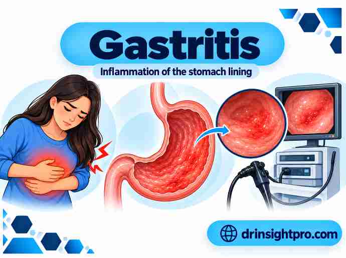
Gastritis is the inflammation of the gastric mucosa (stomach lining). However, it is not a single disease rather, it is a group of conditions with different causes, pathological (tissue) changes and effects on patients. For example, Helicobacter pylori infection causes chronic gastritis, whereas the immune system destroys the stomach glands.
Furthermore, long-lasting inflammation can lead to erosions (shallow sores), ulcers, bleeding, atrophy (thinning of the lining) and in some patients, gastric cancer. The risk depends on the main cause and on whether atrophy or intestinal metaplasia (intestine-like change in the lining) develops.
Types of Gastritis
1. Based on Duration
Acute Gastritis
Acute gastritis is a sudden inflammation of the gastric mucosa with sudden damage to the lining, which may be mild or severe. It can usually heal if the cause is removed early.
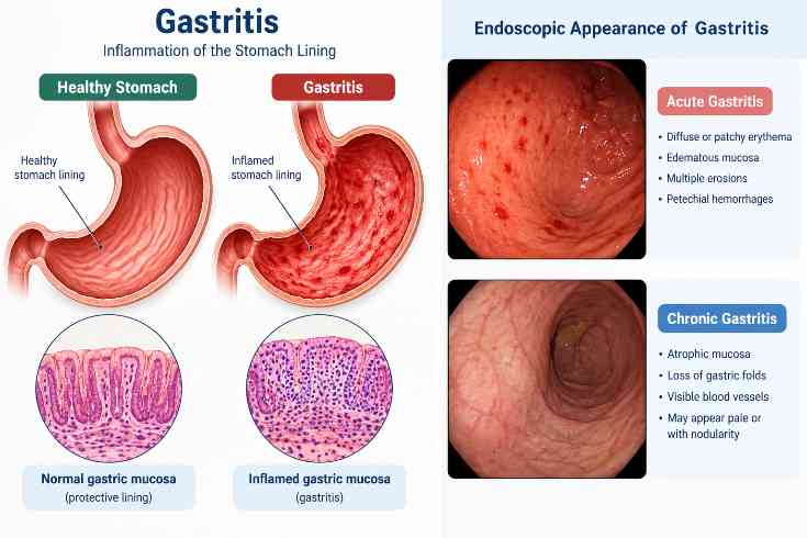
Endoscopic findings (What the camera test shows)
Moreover, endoscopic findings may include:
- Diffuse (spread out) or patchy erythema (redness)
- Edematous (swollen) mucosa
- Multiple erosions (superficial breaks in the mucosa)
- Petechial hemorrhages (small marks caused by bleeding from capillaries)
Histopathology (Findings under the microscope)
In addition, histopathology shows:
- Neutrophilic infiltration (White blood cells in the tissue)
- Surface epithelial damage
- Edema of the lamina propria (swelling in the layer under the surface)
- In severe cases, mucosal necrosis and hemorrhage (death of lining tissue and bleeding)
Chronic Gastritis
Chronic gastritis is a slow, long-lasting swelling of the gastric mucosa and may lasts for months to years.
However, t is most commonly caused by H. pylori infection or by immune attack on the acid-making cells. In contrast, chemical damage is better described as reactive gastropathy (stomach damage without much swelling) because inflammation is minimal.
| Feature | Type A Gastritis (Autoimmune) | Type B Gastritis (H. pylori) | Type C Gastritis (Chemical/Reactive) |
| Definition | Autoimmune destruction of parietal cells and intrinsic factor (a protein that helps absorb B12) associated with antibodies against gastric parietal cells and intrinsic factor. | Caused by infection with H. pylori. | Chemical injury caused mainly by NSAIDs or bile reflux. |
| Common Site | Mostly the gastric body and fundus (main and upper part of the stomach); the antrum (lower stomach is usually not affected) | Usually begins in the antrum; may progress to pangastritis (gastritis of the whole stomach). | Usually the antrum, or diffuse. |
| Endoscopic Features | • Pale, thin lining • Flat lining with fewer folds • Visible blood vessels | • Diffuse erythema • Nodular mucosa (Bumpy lining) • Erosions | • Hyperemia (red mucosa) • Erosions may be present |
| Histology | Chronic inflammation of the body with loss of gland , often with intestinal metaplasia. | Chronic active inflammation (lymphocytes, plasma cells, and neutrophils); H. pylori organisms may be seen. | • Foveolar hyperplasia (overgrowth of surface cells) • Edema • Very few inflammatory cells (key point) |
| Clinical Features | Vitamin B12 deficiency (anemia, neuropathy, glossitis). | Dyspepsia/indigestion and epigastric pain; often asymptomatic. | Dyspepsia and epigastric pain; often asymptomatic. |
| Complications | • Pernicious anemia • Gastric adenocarcinoma (stomach cancer) • Slow-growing stomach tumor | • Peptic/stomach ulcers • Gastric adenocarcinoma • MALT lymphoma (stomach lymphoma (a blood cancer) | • Erosions and bleeding • Ulceration |
2. Based on Endoscopic Appearance (Camera Test)
Erosive Gastritis
Erosive gastritis causes shallow sores in the stomach lining. Although, these sores do not reach the muscle layer.
Non-Erosive Gastritis
Non-erosive gastritis causes no visible sores. Therefore, the lining may look normal. It may also look red or swollen. So doctors often need a tissue sample to confirm it.
3. Types by Causes
- Infection: H. pylori
- Drugs: pain pills called NSAIDs (such as ibuprofen) and aspirin
- Autoimmune: the immune system attacks acid-making cells and causes low vitamin B12
- Chemical damage: bile reflux and NSAIDs. Alcohol can also irritate the lining.
- Stress: very sick patients in intensive care, burns and major injuries
4. Types by Microscope Findings
Doctors can also sort gastritis by the cells they see under a microscope.
- Acute: white blood cells called neutrophils are the main cells.
- Chronic: lymphocytes and plasma cells are also the main cells. The glands may also shrink.
5. Types by Where It Starts
Last, doctors can sort gastritis by the part of the stomach it affects.
- Antral gastritis/Lower stomach gastritis: usually linked to H. pylori.
- Pangastritis/Whole stomach gastritis : affects the entire stomach.
- Body/fundus-predominant gastritis/ Upper and middle stomach gastritis: common in autoimmune gastritis.
How Doctors Rate Gastritis
Long-lasting gastritis can thin the stomach lining. It can also cause intestine-like change in the lining. In addition, rating systems show the risk of stomach cancer. OLGA and OLGIM use the same biopsy plan. It is also called the updated Sydney system. Basically, samples come from the lower stomach (antrum) and the main body (corpus). Therefore they include the bend between them (incisura angularis).
OLGA System
The OLGA system (Operative Link on Gastritis Assessment) uses tissue samples. Meanwhile, it rates how thin the lining is and where. Thinning is the key sign because it links to stomach cancer risk.
- Step 1: Score each area. Therefore, pathologist (lab doctor) scores the thinning in the lower stomach and in the main body:
- 0 = none
- 1 = mild
- 2 = moderate
- 3 = severe
- Step 2: Combine the scores. Moreover, match the scores. Also use the table below. Therefore the two scores give a final stage from 0 to IV.
- Why it matters: Significantly, stages III and IV mean a higher risk of stomach cancer. These patients need regular endoscopy checks.
OLGA list
| Antrum (lower stomach) score \ Corpus (main body) score | 0 | 1 | 2 | 3 |
| 0 | Stage 0 | Stage I | Stage II | Stage II |
| 1 | Stage I | Stage I | Stage II | Stage III |
| 2 | Stage II | Stage II | Stage III | Stage IV |
| 3 | Stage III | Stage III | Stage IV | Stage IV |
OLGIM Staging
OLGIM (Operative Link on Gastric Intestinal Metaplasia Assessment) is like OLGA. It scores intestine-like change in the lining (intestinal metaplasia) instead of thinning.
- Why it is used: Intestine-like change is easier to spot than thinning. So pathologists agree more often. It also helps find patients at risk of stomach cancer.
- Score for each area:
- 0 = none
- 1 = mild
- 2 = moderate
- 3 = severe.
- Final stage: Use the same table as OLGA. Hence, the two scores give a stage from 0 to IV.
- Clinical importance: Stages III and IV link strongly to early cell changes before cancer (dysplasia) and to stomach cancer. So these patients need regular endoscopy checks.
Symptoms of Gastritis
Many patients have no symptoms. When symptoms occur, they often include:
- Epigastric pain/upper belly pain
- Nausea and vomiting
- Bloating and indigestion
- Early satiety/feeling full fast
- Loss of appetite
- Severe cases: vomiting blood, black stools (melena) and tiredness from low blood count
Signs of Gastritis
- Epigastric tenderness/pain when pressing the upper belly
- Pale skin, which may point to ongoing bleeding
Diagnosis of Gastritis
Doctors may also use several tests to check for gastritis and its problems. These include upper endoscopy (a camera test of the stomach) with tissue samples, stool tests, breath tests and blood tests. Therefore, the NIDDK diagnostic guide summarizes these tests.
Laboratory tests:
- CBC: checks for low blood count
- Serum B12: checked when autoimmune gastritis is possible
- Stool occult blood: finds bleeding you cannot see
Tests for H. pylori:
These tests confirm H. pylori infection:
- Urea breath test
- Stool antigen test
- Biopsy urease test/quick tissue test
Upper GI endoscopy:
It shows redness, swelling, shallow sores, bleeding and thinning of the lining.
But the stomach may look normal or unclear. So doctors take tissue samples when needed.
Histopathology/Microscope Tests
A microscope test confirms gastritis and describes it:
- Neutrophils: show sudden or active swelling.
- Lymphocytes and plasma cells: shows long lastingastritis.
The microscope test can also show thinning, intestine-like change in the lining and H. pylori.
Management of Gastritis
Treatment depends on the cause. For example, H. pylori infection needs treatment to kill the germ. Stomach damage from medicines may improve when you use less of them. Therefore, the NIDDK treatment guide gives a patient-friendly overview.
General measures:
- Stop NSAIDs if possible.
- Avoid smoking and alcohol.
- Eat small meals often if this eases symptoms.
Medical treatment:
Acid suppression/to lower acid:
Doctors may give medicines called PPIs (such as omeprazole) to lower acid when needed.
If H. pylori positive:
Furthermore, eradication therapy with a PPI plus antibiotics. For evidence-based regimens, consult the 2024 American College of Gastroenterology guideline on H. pylori treatment.
Autoimmune gastritis:
Patients need vitamin B12 supplements and iron if levels are low.
Severe bleeding:
Doctors treat it during an endoscopy.
Problems Gastritis Can Cause
Gastritis can cause these problems:
- Peptic ulcer disease/stomach ulcer
- Bleeding in the gut
- Iron deficiency anemia/low blood count from low iron
- Pernicious anemia/low blood count from B12 shortage
- Gastric atrophy and intestinal metaplasia/thinning and intestine-like change in the lining
- Higher stomach cancer risk when the lining thins or changes
Latest Research on Gastritis
Research on gastritis is moving fast. Most of it focuses on H. pylori treatment. H. pylori is the most common cause of long-lasting gastritis. Vonoprazan is a newer acid-lowering medicine (a P-CAB). It is now another option next to standard PPIs. The 2024 ACG guideline recommends bismuth four-drug treatment (quadruple therapy) first when antibiotic resistance is unknown. It also lists vonoprazan with amoxicillin as an option.
Similarly, researchers are testing how to make vonoprazan regimens simpler and cheaper. Also a 2026 prospective cohort study examined different vonoprazan dosing strategies in quadruple therapy and reported better cost-effectiveness, which may help in resource-limited settings.
More germs now resist antibiotics. Patients also differ in their genes and sometimes miss doses. These problems have limited standard PPI treatments. So doctors should pick a plan based on local resistance and past antibiotic use. They should also consider each patient. Doctors should also check that the germ is gone after treatment.
Sorting patients by risk is also changing follow-up care. OLGA and OLGIM find patients with advanced thinning or intestine-like change in the lining. These patients need regular checks. Researchers are also testing whether AI can help doctors spot these changes during the camera test. But this tool is still under study. It does not replace a tissue sample. The microscope test is still the best test for staging.
Frequently Asked Questions
Most people with gastritis do not get stomach cancer. But long-lasting gastritis can thin the lining and cause intestine-like change. This is more likely with H. pylori or autoimmune gastritis. These changes raise the risk. OLGA and OLGIM help find patients who need regular checks.
Acute gastritis often heals once the cause is gone. Gastritis from H. pylori can also improve after successful treatment. Autoimmune gastritis lasts for life. But doctors can treat problems like low B12.
Avoid NSAIDs you do not need. Limit alcohol and do not smoke. Treat H. pylori if tests find it. Small meals eaten often may also ease symptoms.
The most common causes are H. pylori infection and long-term use of NSAIDs or aspirin. Autoimmune disease, bile reflux and heavy alcohol use can also cause it. So can severe body stress from serious illness, burns or major injury.
References
- NIDDK. Gastritis and Gastropathy.
- American College of Gastroenterology. Guideline on treatment of H. pylori infection (2024).
- Add primary sources for OLGA/OLGIM (Rugge et al.; Capelle et al.) and the updated Sydney System before publishing.
✍️ About The Author:
Hafsa Hadi is a medical content writer who focuses on gastroenterology. She turns hard research into clear advice for patients.
She writes health guides for medical websites and publications. Clear words matter in patient care. So she writes in a way that is accurate and easy to read. Her goal is to help patients make good choices.
Medical Disclaimer
This article is for education only. Do not treat it as medical advice, a diagnosis or a treatment plan. We aim to share proven medical facts. But health guidelines change often. So this site may not show the latest updates. Medical conditions also differ a lot from person to person. General advice cannot replace a visit with your doctor.
You should always consult a qualified physician or healthcare provider regarding specific symptoms, diagnoses, or treatment plans. Furthermore, never delay or disregard professional medical advice because of something you have read in this publication. If you experience a sudden medical emergency, contact your local emergency services or visit the nearest emergency department immediately.